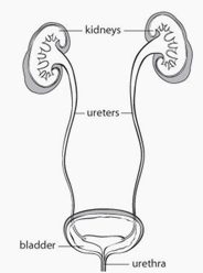
The investigation and treatment of many problems of the kidneys and their drainage system may require visual examination. The ureter is the tube that drains urine from each kidney to the bladder. Ureteroscopy is a procedure in which a narrow scope is passed through the urethra (urinary tube) and bladder, into the ureter toward the kidney.
Ureteroscopy is performed most often for removal of a kidney stone which has become trapped in the ureter. Investigation and treatment of unexplained urinary bleeding or blockage of a ureter may also require ureteroscopy.


Ureteroscopy is performed under anaesthesia. Most cases will require a general anaesthetic (one is “put to sleep”) or spinal anaesthetic (a needle in the back “freezes” you below the waist and you remain conscious). Simpler cases can be performed comfortably with sedation alone.
After the anaesthetic is administered, a long, narrow telescope (ureteroscope) is passed through the urethra into the bladder and up the ureter to the area of concern. X-rays are often taken during the procedure.
 A kidney stone blocking a ureter can often be removed by trapping it in a wire “basket” and carefully pulling it out. Various instruments including laser, ultrasound, or mechanical energy are available to fragment larger stones and allow their passage or removal. These specialized instruments may not be available at all hospitals.
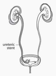
Upon completion of the procedure, a thin plastic tube (ureteric stent) may be placed temporarily in the ureter to prevent blockage while any swelling resolves. This tubemust be removed, usually within a few days or weeks. Many patients can be discharged from hospital on the day of their ureteroscopy.
Urinary infection requiring antibiotic medication is uncommon. Perforation of the ureter may occur. Normally, this will heal rapidly. Rarely, a ureteric injury or abnormal scar formation may require additional surgery to correct the problem.
One should be able to resume all usual activities within a few days.
Burning with urination and blood in the urine are common for a few days after ureteroscopy. This is related to the passage of instruments through the urethra. To help ease these symptoms, drink plenty of fluids and empty your bladder frequently. Small stone fragments may be seen in the urine if the procedure was performed for stone removal.
Pain in the flank or bladder area is common for a several days after ureteroscopy. This pain is usually controllable with a mild painkiller such as acetaminophen (e.g. Tylenol™) or ibuprofen (e.g. Advil™). In some cases, a stronger painkiller available only by prescription, such as acetaminophen with codeine (e.g. Tylenol #3™), may be required. A few patients may have more severe pain or high fever requiring a visit to the emergency room.
Patients requiring a ureteric stent often experience bladder discomfort with increased frequency and urgency of urination. Flank pain with urination or a full bladder, and blood in the urine are not unusual. These symptoms often increase with activity and resolve immediately after the stent is removed.
Some stents have an attached thread that comes out of the urethra. This may be taped to the penis or lower abdomen. We or family physician can remove this stent when appropriate by simply pulling on the thread.
A follow up X-ray is often recommended after stone removal to ensure that there are no residual fragments. If a ureteric stent was placed, you will be notified regarding how and when it will be removed.